Spinal fusion
A spinal fusion is a surgery designed to stop movement in a painful spinal area, and in turn reduce pain generated from the joint. The aim of spinal fusion is to link two individual segments of bone (vertebrae), using metalwork including rods, screws and cages. Sometimes the need for a bone graft is necessary to stimulate bone growth between the vertebrae.
Spinal fusion is used to treat conditions such as spondylolisthesis, scoliosis, fractures and to relieve back and leg pain related to degenerative disc disease (DDD). Once new bone forms, the vertebrae will be linked together, and there should be no further movement between the fused segments. Following surgery, the fusion process usually takes about 3 to 6 months, and can continue for up to 12 months.
Decompression
Decompression refers to any surgical technique that aims to free the space for the nerves in the spinal canal or foramena. A number of different surgical methods are commonly used to achieve a decompression. Depending on what tissue is removed, the procedure may be called: a laminectomy (removal of the bone behind the spinal cord), a foramenotomy (removal of the bone around the spinal nerve) or a discectomy (removal of the spinal disc to relieve pressure).
Laminectomy
A laminectomy is performed to relieve pressure on the spinal cord itself. A laminectomy is most commonly used to treat conditions such as spinal stenosis and spondylolisthesis. This procedure may be performed at the same time as a spinal fusion to prevent instability. In a laminectomy, a small portion of the bone over the nerve root and/or behind the spinal cord is removed. This gives a patient some immediate relief, although residual leg pain, numbness or weakness may continue for several weeks.
Foramenotomy
A foramenotomy is also a type of decompression procedure used to relieve pressure on a nerve. A foramenotomy removes a portion of bone and other tissue that may be compressing the nerve as it exits the spinal column. Most patients obtain immediate relief of their symptoms, and will leave hospital after 2 to 3 days. Some suffer some pain in the neck muscles for a few days and leave hospital between 3 and 7 days.
Microdiscectomy
Microdiscectomy is the current standard surgery for treating a herniated disc (slipped disc / prolapsed disc) and relieving the associated leg pain. Unlike conventional open discectomy, microdiscectomy is performed through a small incision (1 to 1 1/2 inch). The back muscles are lifted and moved away from the spine. After identifying and moving the nerve root, the surgeon removes the injured disc tissue under it.
Effective alternatives to microdiscectomy include:
- Endoscopic microdiscectomy: In an endoscopic microdiscectomy, the surgeon uses a catheter (a thin tube) that contains tiny cameras and surgical instruments through very small incisions. The procedure does not change any of the structural supports of the spine, including joints, ligaments, and muscles.
- Percutaneous discectomy: Percutaneous discectomy (PAD) uses a tube with a device at the tip that cuts away some of the nucleus pulposus at the centre of the disc, and a vacuum that then sucks this gelatinous matter out.
- Laser discectomy: Endoscopic laser foraminoplasty (ELF) uses lasers to locate the likely source of pain. The surgeon can then remove disc tissue, from the centre of the disc, using lasers to burn or evaporate the disc. The incision is tiny and complications are minimal.
Cervical discectomy
Cervical discectomy (neck surgery) is a type of discectomy that can be very effective for relieving the pain associated with compressed nerve roots. In this procedure, the intervertebral disc between two (or more) neck vertebrae is surgically removed. Cervical discectomy involves making a small incision near the front of the neck.
Following removal of the disc, the space between the vertebrae is either left open, or a cage is inserted between the two vertebrae to maintain the normal height of the disc space. Generally, a cervical discectomy is performed at the some time as cervical spine fusion.
X-stop
The X-stop is a titanium implant that is inserted into the spine to relieve painful symptoms of lumbar spinal stenosis. The device is inserted into the lower back at the affected level of the spine. The X-stop is held safely in place behind the spinal cord by wings that prevent it from moving. This implant prevents a patient from bending too far backwards, which can cause leg pain (sciatica) and/or low back pain in patients with sclerosis. This procedure is conducted under minimally invasive surgery, and most patients return home within 24 hours.
Total disc replacement
Total disc replacement is a surgical procedure that is carried out to reduce back pain. The operation involves removing all the damaged disc material in between two vertebrae in the spine and replacing this with an artificial metal and plastic disc. These artificial discs can restore flexibility and reduce the risk of disc degeneration. Disc replacement procedures currently available include: lumbar disc replacement to relieve pain in the lumbar area (lower spine), and cervical disc replacement to relieve pain in the cervix (neck).
The goal is to achieve the same pain reduction as spinal fusion but reduce the limited flexibility of movement that patients experience after spine fusion. The potential advantage of total disc replacement is that the replaced disc would allow motion at the damaged level and would not transfer stresses to adjacent levels.
Disc nucleus replacement
A disc nucleus replacement is a relatively new surgical procedure that aims to alleviate spinal pain. Unlike the total disc replacement, only the inner gel-like core (nucleus) of the disc is removed and replaced with an implant. The outer part of the disc (the annulus) is not replaced.
The implant is an artificial cushioning device called the prosthetic disc nucleus (PDN). A disc nucleus replacement works by restoring the height of the natural disc, which should reduce pressure on the surrounding nerves, thereby relieving pain, numbness and paralysis. The procedure is undertaken using a general anaesthetic.
Kyphoplasty
Kyphoplasty is a minimally invasive surgery that is often used to treat vertebral body fractures, such as compression fractures in the spine. In order to stabilise the crushed vertebrae (bony segments), a balloon is inserted into the vertebral body and gently inflated to restore shape and height to the fractured bone. The balloon is then removed and acrylic bone cement is injected to hold the restored height and shape in place.
Vertebroplasty
Percutaneous vertebroplasty is another type of minimally invasive surgery that orthopaedic surgeons use to treat vertebral fractures in the bony segments of the spine. The surgeon stabilises the fracture with the use of a cement-like substance placed in the space where the fracture has occurred. Vertroplasty is similar to kyphoplasty, but unlike kyphoplasty, no attempt is made to restore the former height of the bone.
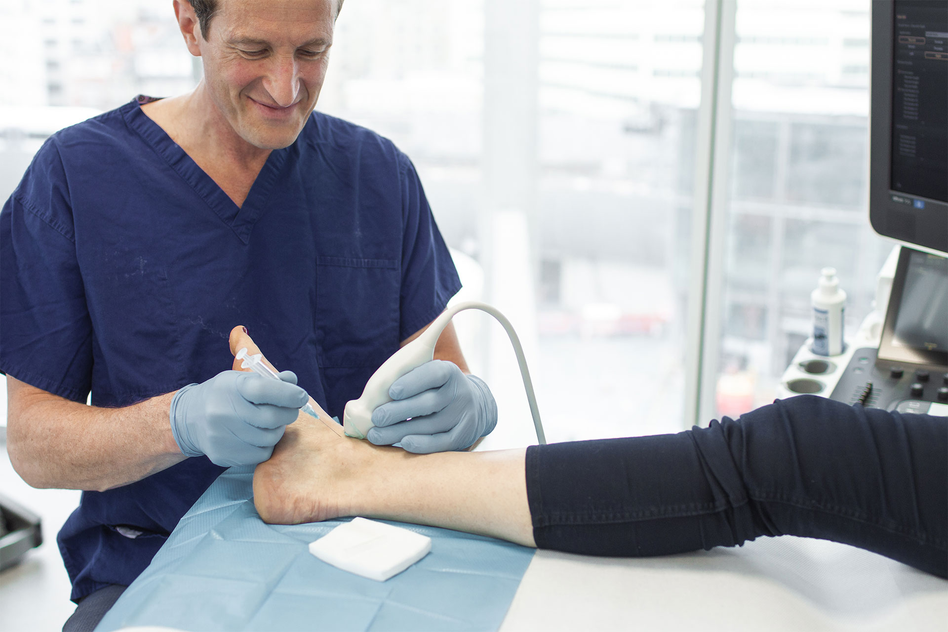
Nerve route injections
Nerve blocks injections are designed to treat chronic and severe back pain due nerve root damage, called radiculopathy. This damage often results from a herniated disc. Nerve blocks can be used to diagnose the exact cause of back or neck pain. They can also be used to treat low back pain and reduce inflammation around the nerve root.
These nerve blocks are injected directly into the exact location of the nerve root, which helps increase the efficacy of the medication. Nerve block injections can avoid the need for surgery if they successfully reduce the pain. The most common type of nerve block injection for nerve root pain is the spinal epidural injection.
Facet joint injections
The facet joints are located on the back of the spine, between each pair of vertebrae. These joints provide stability and give the spine the ability to bend and twist. Facet joint osteoarthritis is caused by the breakdown of the articular cartilage, which can be painful. This can result from previous fractures and injuries but the main cause is spinal degeneration, which occurs slowly over time.
Facet joint injections can be used to diagnose and pinpoint the specific arthritic pain. Facet joint injections can also be used to provide pain relief by injecting anaesthetic and anti inflammatory medications directly into to the area of the arthritis. These injections are performed using X-ray, under a local anaesthetic.
Caudal epidural
In the spine, the membrane that covers the nerve roots is called the dura. The sleeve-like space surrounding the dura is called the epidural space, and nerves travel through the epidural space into the back and legs. Irritation from a damaged disc or contact with the spine will inflame nerve roots in the epidural space, causing pain.
A caudal epidural injection (or ‘caudal’) inserted directly into the epidural space can be used to treat this pain. It uses an anti-inflammatory steroid can block the transmission of signals through nerves in or near the spinal cord. A caudal epidural injection that can cause a loss of sensation and a loss of pain. This can give the inflamed nerve roots time to heal, and provide long-term pain relief. Depending on the success of the caudal, repeat injections may be necessary
Anterior release and fusion
Surgery for scoliosis is indicated when conservative, non-surgical treatments fail. Anterior release and fusion is a surgical technique that can be used to increase the flexibility of spinal curvature. It is indicated in patients with an extremely rigid spinal curve, which is not easily correctable. The anterior release surgery is always followed by a fusion operation to stabilise the spine.
If the scoliosis is in the thoracic (chest) spine, this surgery requires an approach through the chest. Small incisions are made, allowing a surgeon to remove spinal discs in order to loosen up the spine. Screws can then be placed in the vertebral bodies to reduce the curvature of the spine. Additional screws and a rod are inserted to connect the vertebral bodies. This corrects the scoliosis. Bone is added to the disc space to allow the spine to fuse together to provide stability. Compared to posterior release and fusion, anterior release surgery has the advantage of minimal blood loss and less muscle damage.
Posterior release and fusion
Posterior release and fusion is the most commonly used surgical procedure for managing scoliosis. Surgery is made through an incision on the back of and the entire length of the thoracic (chest) spine. The surgeon strips the muscles to the side to allow access to the spine. Hooks and screws are then fixed into the vertebrae. Additional screws and two rods are inserted, one on the left and one on the right, to reduce the amount of spine curvature. This corrects the scoliosis.
Bone is then added to stimulate spine fusion to allow the bones to grow together, and provide stability. Compared with anterior release and fusion, posterior release and fusion usually requires more intensive care and blood transfusions.